Project Sections
- Definition Of Amyotrophic Lateral Sclerosis
- ALS and Cerebellum
- Scope and Aim of the Project
- Chronological Analysis of Clinical Findings
- Visual Analysis of Symptom Progression and Functional Decline
- Early Clinical Findings and Potential Cerebellar Involvement: A Retrospective Analysis
- Clinical Convergence: Aligning Literature Findings with Patient Progression
- Conclusion
- Ethics
Definition Of Amyotrophic Lateral Sclerosis
Amyotrophic Lateral Sclerosis (ALS) is a neurological disease caused by the degeneration of motor neurons in the brain and spinal cord. The causes of the degeneration responsible for the disease include misfolded proteins, impaired RNA metabolism, loss of mitochondrial function, and neuroinflammation. Although research has been conducted on the genetic triggers of this disease, which is not based on a single cause, most cases occur without a family history. The disease has two main types of onset: bulbar and spinal. The bulbar type is the type in which speech and swallowing difficulties begin first. In the spinal type, on the other hand, numbness and loss of strength occur in the arm and leg muscles.
There are two commonly encountered variants of ALS. Primary Lateral Sclerosis (PLS) is a variant that affects only upper motor neurons. It causes spasticity problems that can affect speech, swallowing, and walking abilities. Progressive Muscular Atrophy (PMA) affects only lower motor neurons and causes sudden losses in muscle mass (atrophy). Both variants can progress to ALS over time.
It occurs at a rate of 2–6 cases per 100,000 people each year worldwide. There is no known treatment for the disease. Some medications can be used to prolong the course of the disease. However, these medications may not be suitable for every patient. People with ALS have a life expectancy of 4–6 years after diagnosis. Some patients also live for 10 years or more with good care.
The disease generally presents with physical symptoms. Some of the most common symptoms include:
• Muscle stiffness (spasticity), cramps, or twitching
• Muscle weakness
• Difficulty speaking and swallowing
• Difficulty walking, stumbling, or falling
• Uncontrollable crying or laughing
• Fatigue
• Difficulty breathing
The symptoms are not sufficient to diagnose the disease. Some tests are performed to make an accurate diagnosis or to distinguish diseases that resemble ALS. The disease can be diagnosed through reflex tests, laboratory tests, biopsy, MRI, and EMG.
Since ALS is a disease that affects motor neurons, it affects physiological processes that can be consciously controlled. However, cognitive processes continue in most patients unless they have a coexisting disease such as FTD-ALS. Death usually occurs due to respiratory difficulties.
ALS and Cerebellum
Historically, the currently accepted view of ALS has been that it does not cause any damage to the cerebellum. However, with the development of medical imaging and testing methods over time, studies have been conducted suggesting that the cerebellum may also be affected by this degenerative process. These discussions are still ongoing today. Neuroimaging studies and neuropathological findings have provided information indicating degeneration in the cerebellum. Changes in the proportions of gray and white matter in the cerebellum have been recorded. ALS is thought to affect not only the upper and lower motor neurons but also the areas connected to them. Research is ongoing on the multisystemic nature of ALS. Some studies suggest that a part of the cerebellum or the region above the cerebellar tentorium, known as the supratentorial region, may be affected.
As research continues, there is still no clear consensus on this issue. However, some studies suggest that certain symptoms may be indirectly related to this.
Scope and Aim of the Project
What Does the Project Examine?
In this project, more than two years of a patient’s medical data were examined chronologically. In addition, the patient’s physical symptoms during this period were observed and documented. The aim was to present the rate of disease progression and the physical changes occurring during the course of the disease based on the data. The data examined include reflex tests, biopsy results, laboratory tests, EMG, and CT data. The changes that occurred were shown by comparing monthly periods with the dates recorded in hospital reports. Some diagnostic processes and findings considered potentially relevant were added to the chronology.
What Does the Project Aim to Do?
The project aims to transparently show the course of the disease on a monthly basis, both medically and physically. It provides a chronology of disease progression in a disease that does not follow a predictable course. It also examines the possible connection between some of the patient’s diagnoses mentioned in the data and the cerebellum. The project also explores whether some earlier findings in the patient’s records may have been relevant to the later diagnosis. This observation is based on a single case and does not establish a medical conclusion.
What Are the Project’s Sources and Limitations?
The project presents the processes transparently. The differences found between the medical data were objectively compiled from the patient’s medical reports. During the reporting of physical symptoms, observations were made firsthand and care was taken to report them objectively. The tests and reports were obtained from official medical records. In addition, actual dates were used, and no changes were made to the dates. The patient’s diagnoses and treatments during the process are clearly stated. However, this project has not received any official approval or undergone peer review. Given the educational level of the person who prepared the project, it does not constitute a medical recommendation or indicator. Since only one patient’s data were examined, the project does not guarantee that the disease will progress in the same way in every patient. The person who prepared the project does not have an undergraduate education. None of the information in this project should be considered medical advice.
Chronological Analysis of Clinical Findings
This section examines the patient’s medical records in chronological order. It focuses on how symptoms, test findings, diagnoses, and treatment changed over time.
23.05.2022
In May 2022, the patient went to the hospital due to sudden-onset dizziness, nausea, and loss of balance. The patient’s medical history was taken and a physical examination was performed by an ENT specialist. The patient’s medical history showed no evidence of physical trauma. The physical examination performed by the doctor did not reveal sufficient findings for a diagnosis of vertigo. However, considering the patient’s clinical symptoms, a diagnosis of vertigo was made, and treatment was then started.
08.06.2022
During this period, the patient continued to experience severe dizziness and loss of balance despite the ongoing treatment. Due to the patient’s ongoing complaints, the patient went to the neurology outpatient clinic. After the examination, the neurologist made the same diagnosis and recommended continuing the treatment.
27.10.2022
Over the following approximately four months, the patient did not get the expected benefit from the treatment and went to the ENT outpatient clinic again. The diagnosis was maintained and the treatment was continued.
06.06.2023
During this period, the patient continued to experience loss of balance. Over this period, the patient also began to experience weakness in the hand and slowed speech. The patient began to drop objects from time to time due to the loss of strength. For this reason, the patient went to the neurology outpatient clinic. EMG, brain CT, and blood tests were requested for the patient.
The EMG results led to a diagnosis of carpal tunnel syndrome.
The non-contrast brain CT showed the following findings. The bone structures included in the examination are intact. The cerebellum, fourth ventricle, and brainstem appear normal. There is enlargement of the third ventricle, lateral ventricles, basal cisterns, and hemispheric cortical sulci due to atrophy. The bilateral thalami, internal capsule, basal ganglia, and centrum semiovale are normal. After reviewing the non-contrast brain CT results, the doctor requested contrast-enhanced carotid artery CT angiography.
The contrast-enhanced carotid artery CT angiography showed the following findings. No pathological findings were detected in the aortic arch or at the origins of its major branches. Both common carotid arteries and the bifurcation levels appear normal. No pathological findings were detected in the cervical, petrous, precavernous, cavernous, and supraclinoid segments of the internal carotid arteries.
As a result of all these tests, the neurologist could not make a definitive diagnosis but considered the possibility of ALS.
07.08.2023
Over this period, the patient continued to experience increasing loss of strength and balance. The speech disorder began to become more noticeable. The patient went to the same neurologist for an examination. The non-contrast brain CT requested by the doctor showed similar findings. The bone structures included in the examination are intact. The cerebellum, fourth ventricle, and brainstem appear normal. There is enlargement of the third ventricle, lateral ventricles, basal cisterns, and hemispheric cortical sulci due to atrophy. The bilateral thalami, internal capsule, basal ganglia, and centrum semiovale are normal. No intra- or extra-axial hemorrhage or midline shift was detected. A midline, approximately 5 × 11 mm lipomatous hypodense lesion with fat density was observed, extending from the level of the atrium of the lateral ventricles to the interhemispheric fissure. As a result, the doctor referred the patient to a hospital with more advanced facilities.
11.09.2023
Over the course of approximately one month, the patient’s speech impairment became rapidly more noticeable. In addition, weakness in the hand began to significantly affect daily life, while the loss of balance continued. At the hospital the patient was referred to, the neurologist requested a non-contrast brain CT. In the posterior fossa, the densities of the cerebellum and brainstem are normal. The basal cisterns are patent. The fourth ventricle is in the midline and is normal in size. In the supratentorial sections, the gray and white matter densities, sulci, and gyri of both cerebral hemispheres are normal. No midline shift was observed. The ventricular system is normal in size. The sella, parasellar structures, and both orbits included in the examination are normal. As a result, the doctor diagnosed the patient with vertigo. This was the last diagnosis of vertigo the patient received.
Due to the loss of strength in the hand, the patient was referred to physical therapy and treatment was started.
22.09.2023
The doctor re-evaluated the patient based on the previous test results. The patient was placed under follow-up for a possible diagnosis of ALS.
03.10.2023
The patient went to the orthopedic outpatient clinic due to weakness and pain in the left foot. However, no diagnosis was made and no treatment was started.
22.11.2023
During the following two months, the doctor followed up with the patient. The patient’s complaints continued to worsen. The non-contrast brain CT performed on this date showed the following findings. In the posterior fossa, the densities of the cerebellum and brainstem are normal. The basal cisterns are patent. The fourth ventricle is in the midline and is normal in size. In the supratentorial sections, a decrease in the white matter density of both cerebral hemispheres is observed. The supratentorial ventricles, cerebral sulci, and fissures appear wider than normal. No midline shift was observed. The sella, parasellar structures, and both orbits included in the examination are normal.
16.02.2024
Speech has slowed significantly, and swallowing problems have started. The weakness in one hand has made it impossible to use that hand. The weakness that started in the left foot has worsened the balance disorder. As a result, the patient went to the hospital again and underwent a non-contrast brain CT examination. Some differences can be seen in the brain CT results compared with the previous examination. No changes were detected in the cortical areas of both cerebral hemispheres or in the subcortical and deep white matter density. The bilateral basal ganglia, thalami, internal and external capsules are normal. The third, fourth, and both lateral ventricles are normal in size. There is a pericallosal lipoma extending to the atrium of the lateral ventricle.
Although the disease progressed rapidly physically over the approximately three-month period, there are some differences between the two brain CT examinations. Compared with the previous report, which stated that there was a decrease in white matter density, no changes were reported in the brain CT performed in February. Similarly, while the previous brain CT stated that the cerebral sulci and fissures were wider than normal, the brain CT performed in February reported that both lateral ventricles were normal in size. This suggests that although the disease progressed physically during the follow-up period, this progression may not appear to the same extent in brain CT results. Therefore, when evaluating the clinical progression of ALS, it is also important to consider the patient’s physical and clinical condition, rather than relying only on brain CT findings. During this period, when the physical manifestations of the disease became more noticeable in the patient, the differences between the brain CT results suggest that the disease may have physical effects even when visible progression is not always seen in test results.
08.03.2024
As the patient’s existing problems worsened and the swallowing difficulty became more noticeable and began to affect daily life, the patient went to the neurology outpatient clinic again. The same doctor, who had been following the patient for a long time, requested a non-contrast brain CT and carotid artery ultrasound. In the posterior fossa sections, the cerebellar hemispheres and brainstem are normal. The fourth ventricle is in the normal location and has a normal configuration. The basal cisterns are normal in size. No space-occupying lesion was detected in the posterior fossa. In the evaluation of the supratentorial sections, there is no pathological change that can be identified as an intracerebral hypo- or hyperdensity. The third ventricle is in the midline and is normal in size. No displacement of the cerebral midline structures was detected. The cerebral sulci are normal in size.
There is a minimal increase in intima-media wall thickness in the bilateral carotid vascular structures. A minimal atheromatous plaque was observed at the level of the bulb of both CCAs. Apart from this, the caliber and walls of the bilateral carotid and vertebral arteries are normal. On spectral examination, the flow velocities, directions, and patterns of the bilateral CCA, ECA, ICA, and vertebral arteries are normal.
Although sufficient data were not available to establish a diagnosis in the patient, who had been followed for a long time, the patient’s clinical progression closely matches the progression of ALS. However, since a diagnosis could not be established, the process continued and no treatment was given. As a result, the doctor referred the patient to a higher-level medical center.
Despite the lack of sufficient data in the tests performed during the patient’s long-term follow-up, the disease progressed in a clinical course similar to that of ALS. The expected course of the disease could not be observed in the tests performed to identify the disease. It is known that the disease can progress differently in each patient. In addition, differences in disease progression may have certain consequences, one example being that the progression of the disease may not always be regularly observable. For this reason, differences in disease progression can significantly prolong the diagnostic process. An example of this can be seen in this patient: despite the disease having progressed considerably, a diagnosis could not be established.
20.04.2024
The patient’s speech problems became particularly noticeable and, together with the swallowing problems, became the most visible functional loss during this period. At the same time, weakness in the other hand progressed rapidly. Loss of balance also became more frequent due to weakness in the foot. In addition to the ongoing complaints, inappropriate laughing and crying began. At the higher-level medical center the patient was referred to by the doctor, a needle EMG and a biopsy were performed. During the needle EMG, the cervical, thoracic, lumbosacral, and bulbar regions were examined. Electrophysiological findings consistent with carpal tunnel syndrome were detected in the left hand. Apart from chronic neurogenic MUP changes accompanied by mild denervation in the dorsal interossei, no significant pathological findings were detected in the other muscles examined. The needle EMG was interpreted as largely normal and was reported as such.
A biopsy was performed using a tissue sample taken from the patient’s left ankle. However, no conclusive result was obtained.
During this process, doctors discussed many diseases that could mimic ALS or have a similar course, but these possibilities were ultimately ruled out. The fact that the needle EMG did not largely produce the expected result suggests that the disease cannot be diagnosed using only one standard test. For this reason, performing different tests and following the patient’s clinical course over a long period are important for the diagnosis of ALS. Similarly, ruling out diseases that mimic or resemble ALS also plays an important role in this process.
The patient was referred to another doctor who specializes in ALS for further diagnosis.
08.05.2024
A PET-CT scan was performed at the request of the specialist the patient was referred to, as the patient’s existing complaints continued to worsen. A PET-CT scan showed the following findings. In the head and neck region, symmetric F-18 FDG uptake was observed in the bilateral palatine tonsils. They were initially considered to be possibly secondary to reactive processes. A lymph node in the right cervical level 2A region showed mildly increased F-18 FDG uptake. F-18 FDG uptake in the head and neck region was within physiological limits. In the thoracic region, F-18 FDG uptake in both lung parenchyma and the mediastinum was within physiological limits. In the abdominal region, mildly increased diffuse F-18 FDG uptake was observed in the stomach. F-18 FDG uptake in the liver, spleen, and other abdominal organs was within physiological limits. In the pelvic region, F-18 FDG uptake in the pelvic organs was within physiological limits. In the skeletal system, mildly increased F-18 FDG uptake was observed in subcutaneous nodular lesions in the posterior aspect of both humeri. Increased F-18 FDG uptake was observed in the area corresponding to the left intercondylar fossa and around the left shoulder joint. They were initially considered to be possibly secondary to degenerative processes. Focal increased F-18 FDG uptake was observed in the muscle tissue adjacent to the distal right tibia. An appearance consistent with thoracotomy was noted in the sternum.
F-18 FDG uptake on PET-CT does not always indicate a disease on its own; there may also be infection-related cells with increased metabolic activity in areas of uptake. In addition, the PET-CT was performed because the patient’s other test results did not clearly indicate ALS. This may make it possible to rule out another possible disease, such as paraneoplastic neurological syndrome (PNS). As a result, the possibility of another disease was ruled out. It may be considered that the disease could have a course that is not detectable through the patient’s test results.
24.05.2024 – 30.05.2024
In this period, the patient was admitted to the hospital for an official diagnosis. During this period, the patient’s swallowing problems progressed, and the patient began to have difficulty even when drinking water. The patient’s speech became unintelligible, and the patient began to need support while walking. Due to weakness in the hands, the patient is unable to perform many daily activities. A needle EMG was performed, but no finding directly indicating ALS was identified. The patient’s neurological examination and muscle strength examination were performed during the hospital stay. During approximately one week of follow-up in the hospital, the patient’s previous test results were also reviewed. The decision was made by considering the patient’s current condition and the clinical course of the disease throughout the long-term follow-up. The patient was diagnosed with ALS and discharged from the hospital.
30.05.2024 – Present (2026)
Since the diagnosis was made, the disease has progressed rapidly. The patient’s swallowing difficulty has become rapidly more noticeable, and the patient aspirates especially when consuming liquids. For this reason, a PEG was placed. Due to loss of strength in the hands and arms, the patient has become unable to perform daily activities. The patient, who was previously able to walk with support, first became unable to walk and later became unable to sit without support. At this point, the patient needs the care of another person to continue daily life.
Visual Analysis of Symptom Progression and Functional Decline
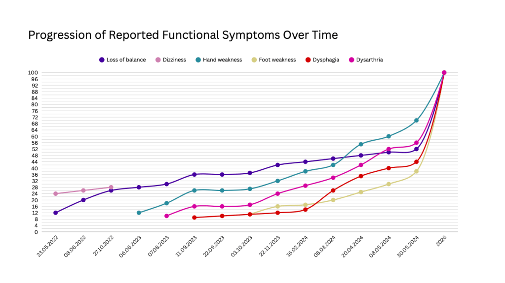
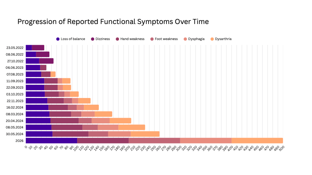
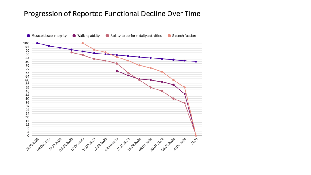
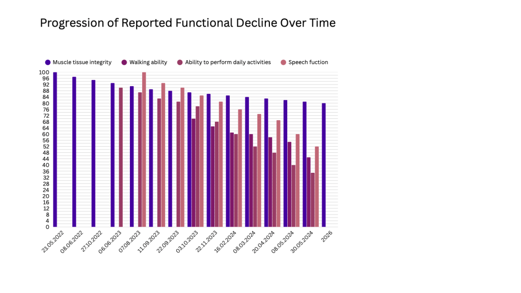
This section visualizes symptom progression and functional decline over the roughly two-year observation period through graphs. The values are not based on exact mathematical calculations, but rather on the visibility of progression during the observation process. As an exception, muscle tissue loss was calculated using actual measurement values.
Early Clinical Findings and Potential Cerebellar Involvement: A Retrospective Analysis
This section of the study presents a retrospective analysis based on research into the effects of ALS on the cerebellum, which has been a topic of interest in recent years. In this process, it puts forward a proposition about whether the patient may have had an early sign of the disease, based on vertigo and the basic functions of the cerebellum. However, since this is a patient-specific evaluation, it does not express a general conclusion.
The cerebellum is located behind the brainstem and is in close proximity to the pons. The fourth ventricle is located between the brainstem and the cerebellum. The basic functions of the cerebellum include maintaining balance, motor coordination, muscle tone, and spatial perception. Since ALS is a disease that primarily affects the upper and lower motor neurons, there is no evidence of a direct relationship between ALS and the cerebellum. However, in recent years, research has been conducted into this possible interaction through advances in neuroimaging.
Vertigo can occur due to different causes and can basically be divided into two groups. Peripheral vertigo originates from the inner ear or the vestibular nerve, and one of its most common causes is BPPV. Central vertigo originates from structures associated with the vestibular system in the brainstem, cerebellum, or other parts of the brain. The patient was told that the diagnosed peripheral vertigo was caused by BPPV. Benign Paroxysmal Positional Vertigo (BPPV) occurs when otoliths in the inner ear become displaced from their normal position and enter the semicircular canals. Factors that may trigger BPPV include aging, head trauma, prolonged immobility, inner ear diseases, or migraine. It is known that the patient had not experienced any of these factors before the onset of vertigo. Furthermore, the ENT specialist stated that the physical examination did not reveal any findings that could cause BPPV. However, considering the patient’s clinical course, the doctor diagnosed the patient with peripheral vertigo.
As can be seen from a retrospective evaluation, there is only a limited period between the patient being diagnosed with vertigo and ALS beginning to be recognized. The diagnosis of vertigo was also repeated after the patient developed weakness in the hand and speech problems.
Based on this, the possibility that the patient was experiencing central rather than peripheral vertigo can be considered. In this case, if it is assumed that ALS has an effect on the cerebellum, the central vertigo that developed in the patient could be explained. However, since this has not been proven, this is only a proposition. Although it is possible to make this connection retrospectively, it cannot be said that an earlier diagnosis was possible, since this type of inference could not have been made at the beginning of the process.
Nevertheless, in the summary of the proposition, it states that the vertigo experienced by the patient may have been central vertigo originating from the cerebellum, based on the cerebellum being the center of balance and coordination. It questions whether the involvement of the cerebellum could be a result of ALS.
The variable nature of ALS and the fact that the relationship between ALS and the cerebellum has not been proven should be taken into consideration. In addition, the possibility of peripheral vertigo occurring due to age should also be considered, since the patient’s age is consistent with this factor.
Clinical Convergence: Aligning Literature Findings with Patient Progression
This section of the study evaluates how the general views about ALS in the literature apply to the patient and whether the patient’s clinical course is consistent with the literature. In making this evaluation, it was taken into consideration that the disease may not always progress in the same way.
The general patterns of ALS onset usually include loss of balance, weakness, and slowed speech. These problems appeared in the patient over time in a way that was consistent with the general description of the disease. In terms of progression, the symptoms increased over time and followed a course that was consistent with the general progression of the disease.
There is no specific test that directly identifies ALS. Instead, the diagnosis is made by ruling out similar diseases and through clinical observation. CT, EMG, PET-CT, and similar tests were performed on the patient many times over time to rule out other diseases and identify the disease. However, these tests did not show any clear findings indicating ALS. An MRI could not be performed because the patient has a mechanical heart valve that was implanted approximately 25 years ago. Although the diagnosis process took a long time, the diagnosis was made by taking the clinical course into consideration. As a result, it is not possible to say that the patient was fully consistent with the literature in this regard.
When it comes to the stages of ALS and the rate of progression, however, the patient shows a similar pattern. It took several years for the disease to reach advanced stages. Although the disease sometimes progressed more rapidly during this period, it generally followed a similar course. In this respect, it is possible to say that the patient was consistent with the literature.
Conclusion
In conclusion, throughout the study, information was provided about the definition of ALS in the literature and research on ALS and the cerebellum. Approximately two years of data were examined through analysis, reported transparently, and supported by graphs. A proposition based on the possible connection between ALS and the cerebellum was presented, and the patient’s consistency with the literature was analyzed.
The study was based on information that was current as of 2026.
Ethics
The study contains detailed information, and for this reason, certain ethical considerations were taken into account throughout the study. Permission was obtained from the patient mentioned in the report to use the patient’s data. No information that could compromise the patient’s privacy is included in the study. No hospital, doctor, or medication names are given in the study. There are no identifying or accusatory elements in the study. The scope and purpose of the project are clearly stated in the introduction. It is clearly stated that the project does not constitute medical advice. No information was changed in order to maintain the transparency of the project. All dates and tests were compiled from official records.

